Innovation in the National Veterinary Schools of France (ENVF): a virtual microscope offers a new educational dimension.
Led by the École Nationale vétérinaire de Toulouse (ENVT), the PATIM project ("Plateforme d’Apprentissage Transdisciplinaire par l’Image Numérique au service de la formation clinique vétérinaire", which can be translated in "Transdisciplinary Learning Platform by Digital Image at the service of veterinary clinical training") of digital and technological innovation of the image is being deployed in the four veterinary schools in France (Alfort, Lyon, Nantes, Toulouse) [in French].
Students will be able to benefit from a virtual microscope as an educational tool to reinforce the acquisition of fundamental and clinical knowledge essential in the diagnostic process. They will be able to observe scanned tissue slides on their screen. A connection to a single platform that can be done from anywhere, at any time and accessible as simply as a connection to a website.
The microscope is the tool par excellence for analyzing the structure, constitution, and also the dysfunctions of a tissue. Current practices have always required the use of an optical microscope to visualize glass slides on which stained tissue preparations have been deposited. This visual recognition and learning step is an important part of the training of future veterinarians.
The digital transition, which is essential today and applied to almost all fields, has also been applied to the microscope: the image that was observed through the microscope can now be digitized and observed on a computer screen. Although this digital dimension offers considerable advantages, the very large size of the images (up to 150 gigabytes per image), the multitude of formats and the IT infrastructure to be deployed to access them have long been an obstacle to the sharing of images and their use in an educational component.
It is in this context that the PATIM project "Transdisciplinary Learning Platform by Digital Image at the service of clinical veterinary training", itself integrated into the program "Pedagogical innovation in agricultural and veterinary higher education" implemented by the Agronomic, Veterinary and Forestry Institute of France (Agreenium), was created.
« The objective of the PATIM project is to use the educational innovation potential of digital and technological developments in the image, for the benefit of veterinary education. The purpose of this platform is to facilitate and strengthen the acquisition, by the student, of fundamental and clinical knowledge, but also of analytical skills and diagnostic approach, through practice in a playful and interactive virtual situation », specifies Mrs. Raymond Letron, professor of histology and veterinary pathological anatomy, coordinator and leader of the PATIM project at ENVT.
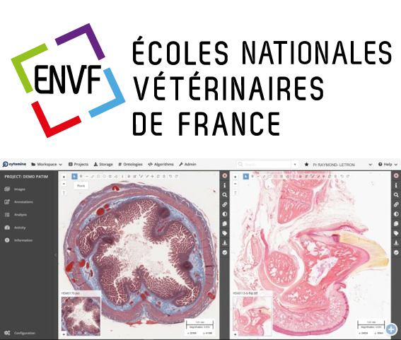
The CYTOMINE web interface makes it possible to share very high resolution images online and offers the same navigation parameters in the tissue as the magnifications of a microscope. CYTOMINE also offers the advantages of being able to annotate images and share content, strengthening collaborative work and offering a new educational dimension to future veterinarians in training.
The start-up CYTOMINE which developed this virtual microscope is based in Liège in Belgium, and is delighted with the initiation of the PATIM project on this platform.
NB : the images used in the illustration above can be explored using Cytomine by following the following links: